Welcome to the OHAGAN LAB at Montclair State UniversityHow is microtubule-based transport of cargo regulated in neurons?
Motor molecules such as kinesins and dyneins, acting as cargo trucks, travel on microtubule highways to deliver cargos inside cells. But how are cargos delivered to the right place, and in the right amount, inside cells? We are a genetics and cell biology lab that uses a microscopic roundworm called C. elegans to study “signposts” on microtubule "highways" inside neurons. The Tubulin Code Hypothesis suggests that microtubules incorporate different tubulin isoforms and post-translational modifications (PTMs) that convey information, like "signposts," to different molecules that interact with microtubules, such as motor molecules. For example, glutamylation side-chains can be reversibly added to microtubules to regulate both the structure of the microtubule highways, as well as traffic carried by motors. MAPs (microtubule-associated proteins) also regulate microtubules and motor molecules. The Tubulin Code might also regulate binding of MAPs to microtubules. We are currently using genetics methods to determine how localization of ciliary MAPs respond to differences in the Tubulin Code, including microtubule glutamylation and tubulin isoforms. We are also interested in how microtubule glutamylation might be regulated by the presence or absence of particular MAPs. Together, these two regulatory layers may be essential for the function and health of neurons, as defects in the the Tubulin Code and the function of MAPs, and resulting dysregulation of microtubule-based transport in neurons, are associated with neurodegenerative diseases. |
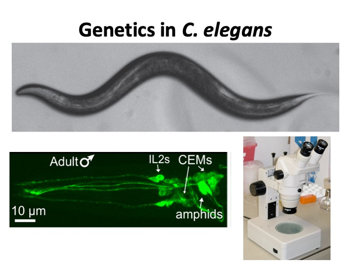
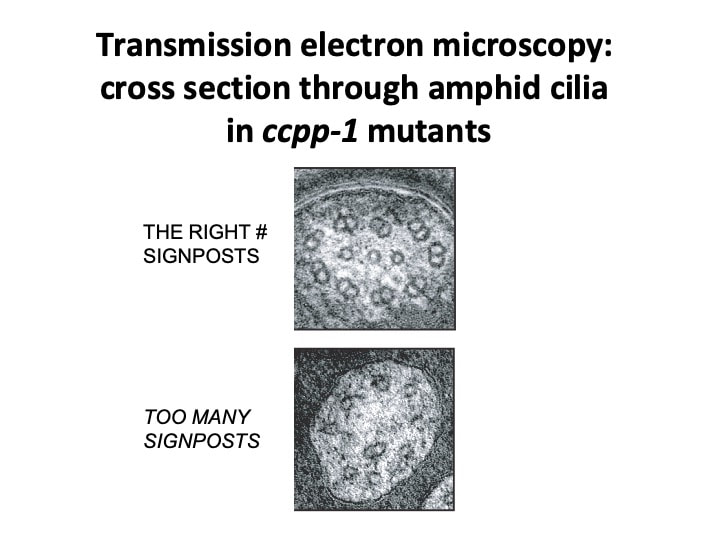
We are using genetic methods in C. elegans and fluorescence microscopy to detect differences in subcellular localization of fluorescently-tagged MAP genes when particular tubulin genes or enzymes that regulate microtubule glutamylation are defective. The nervous system of C. elegans is well characterized and the entire connectome of the 302 neurons present in hermaphrodites has been reconstructed using electron microscopy. Because C. elegans is transparent, it is easy to visualize individual identified neurons expressing GFP markers. We also use established behavioral assays to assess the function of particular neurons. We are most interested in ciliated sensory neurons. Above right, Transmission Electron Microscopy allowed us to look directly at the structure of the microtubule cytoskeleton in sensory cilia. The micrographs shown are cross-sections through a single sensory cilium in wild type (top), in which the right number of glutamylation "signposts" are present, and in a ccpp-1 mutant, in which there is excessive hyperglutamylation--too many glutamylation "signposts," resulting in defects in the structure of ciliary microtubule doublets.
